View Profile

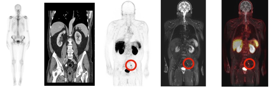
Molecular imaging uses small quantities of radioactive tracers to image the body at the molecular and cellular level. These tracers show how the body is functioning and enables measurement of chemical and biological activities in the body. By using specific tracers, radiologists can image processes within the body to add to the information that anatomical imaging such as CT or MRI provide. For example, a tracer called FDG, which is similar to glucose (sugar) can map areas that utilize high levels of sugar (high metabolism), such as a tumour – while others can be designed to bind to specific proteins or targets (receptors) on cells.
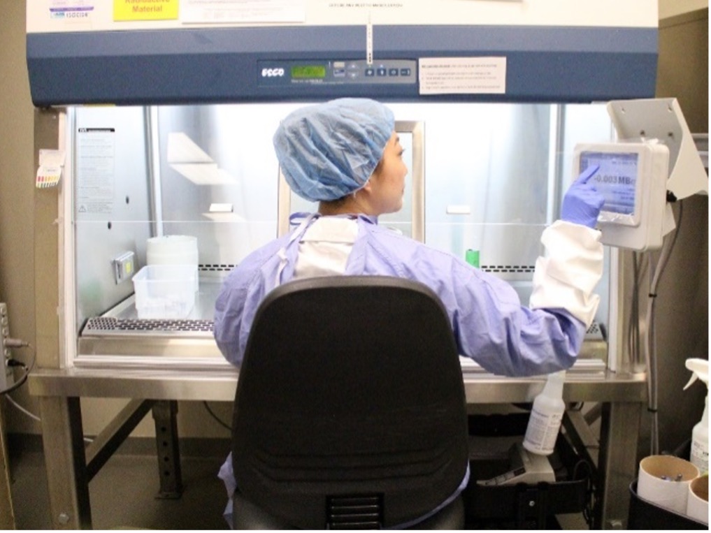
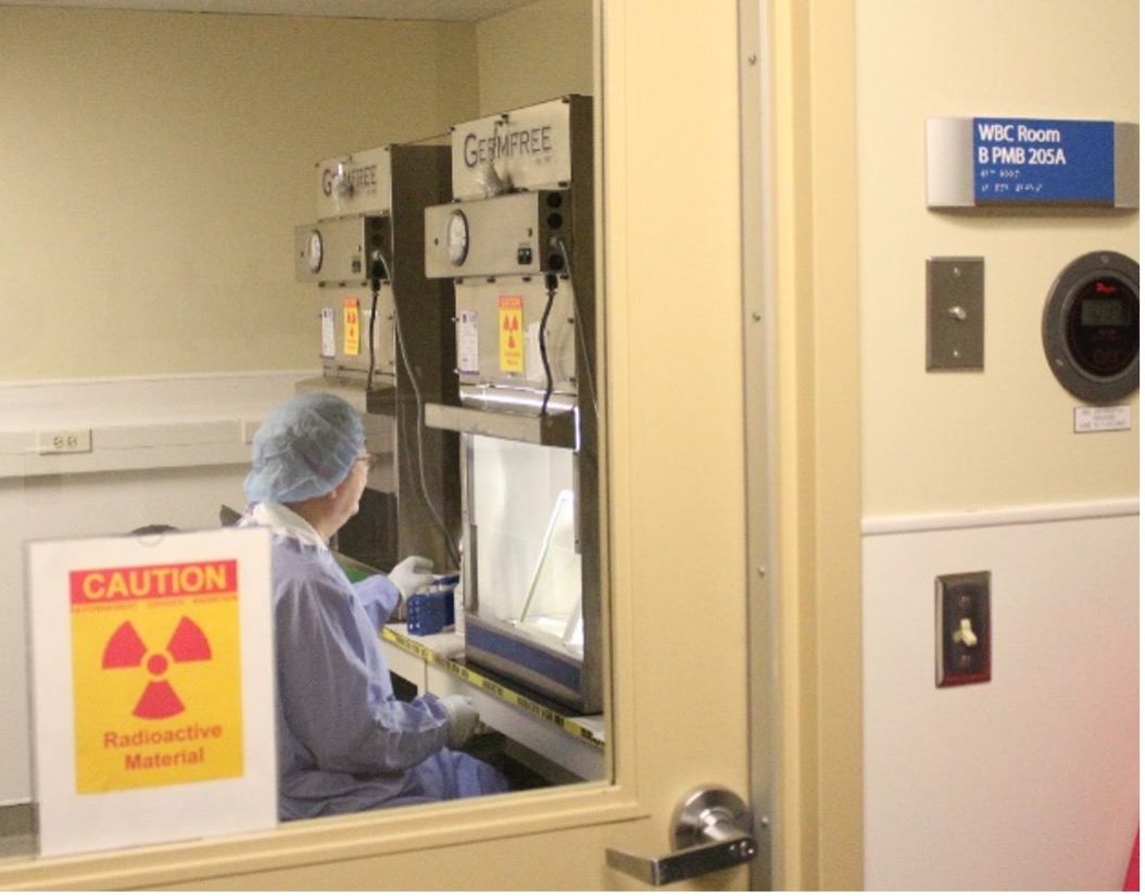
The UHN Radiopharmacy manufactures, tests and packages radioactive pharmaceuticals used in Nuclear Medicine studies. Our facility holds a Drug Establishment License from Health Canada and our products are manufactured according to good manufacturing practices (GMP).
Some of our services provided on both a routine and emergency basis include:
The Cylotron facility is managed by Canprobe, a joint venture between UHN and the Centre for Probe Development and Commenrcialization in Hamilton to create a Canadian Centre of Excellence for the development, translation, utilization and commercialization of molecular imaging probes. CanProbe leverages the strengths of its partners and affiliates.
There are multiple gamma cameras and SPECT/CT scanners installed across the various sites.
The Division has 2 PET/CT scanners including a recently installed state-of the art digital PET/CT scanner (Siemens Biograph VISION) at the Princess Margaret Cancer Centre and a 3 Tesla PET/MR system (Siemens, mMR) at Toronto General Hospital.
Over 2300 general nuclear procedures are performed annually across all University Health Network, Sinai Health & Women’s College Hospital sites including bone scintigraphy, 14C breath test, V/Q scans, sentinel lymph node mapping, radionuclide angiography, myocardial perfusion imaging amongst others.
There are over 4400 PET/CT scans performed annually. The clinical PET program is a dynamic and growing program, with an approximately 10% increase in volumes annually. The vast majority of PET scans are for oncology indications with FDG, 68Ga-DOTATATE for neuroendocrine tumor imaging, 18F-DCFPyL (PSMA) for prostate cancer imaging as well as other novel tracers through various clinical trials.
82Rb PET for myocardial perfusion imaging is planned for a dedicated PET/CT scanner located at the Toronto Western Hospital (Nuclear Cardiology, 5th floor).
Theranostics is an emerging branch in cancer diagnosis and treatment incorporating therapeutics with diagnostics (=theranostics), which heavily leverages the expertise in JDMI’s Molecular Imaging Division. The radiopharmaceuticals made for theranostics can function in multiple roles, with the radiation emitted used for controlling cancer cells as well as for imaging. Theranotics enable targeted personalized therapy, often for difficult to treat cancers, and have been shown to improve quality of life. The duality of these agents also represents a convergence of sciences: chemistry, pharmaceutics, imaging, radioisotope generation, nuclear medicine, radiation and medical oncology and clinical outcome measurement.
The concept of theranostics has been around for many decades with 131I therapy for thyroid cancer (with over 150 patients treated annually at UHN) but has evolved to include other radiopharmaceuticals including 223Ra dichloride therapy (Xofigo) for metastatic prostate cancer (approximately 140 doses administered annually), and 177Lu DOTATATE therapy for metastatic neuroendocrine cancers. In the coming year, UHN will also be collaborating with industry to conduct a clinical trial of 177Lu PSMA therapy for patients with metastatic castration-resistant prostate cancer after second line hormonal therapy. The existing clinical expertise, and research and innovation ecosystem at UHN puts us in a unique position to launch novel theranostic radiopharmaceutical trials in collaboration with Canprobe, the Centre for Probe Development and Commercialization and industry partners.
The Division is actively involved in training of medical students, residents and fellows including 1-2 clinical-research Molecular Imaging fellows annually. There are also multiple rotating residents and fellows from other Divisions within JDMI. In 2021, a unique University of Toronto fellowship in Molecular Oncology Imaging and Therapeutics will be launched. This cross-department fellowship is a 2-year clinical research fellowship in medical imaging and radiation oncology at the University Health Network.
The existing infrastructure provides a fertile ground for research led by the Division’s staff and trainees. Two of our staff have protected research time through the JDMI Research Leadership awards. Between January 2018 and December 2020, members of the division have published over 70 manuscripts in peer reviewed journals and have presented their work in the leading society meetings (Society of Nuclear Medicine & Molecular Imaging, European Society of Nuclear Medicine, Radiological Society of North America, and the Americam Roentgen Ray Society annual meetings among others.
There are currently > 30 research projects ongoing in Molecular Imaging. Research projects in PET/CT and PET/MR span wide areas in imaging-based oncology research including prostate cancer, sarcomas, lung cancer, breast cancer, liver cancer, esophageal cancer, H&N as well as gynecological diseases. Furthermore, non-oncological indications like transplant graft evaluation, peripheral artery disease and rheumatoid arthritis are being investigated. Cardio-vascular research, dedicated MSK projects and also an array of PET-based and MR-based technical research is being conducted in the context of hybrid imaging. Molecular Imaging has well established co-operations on different levels of research directly with headquarters of the vendors were are working with. Consecutively, joint projects are being carried out continuously and the division has a successful track record of attracting industry as well as competitive research funding.
6 Staff Radiologists including 4 staff with cross appointments in other division, enhancing the multidisciplinary expertise across the division. Trainees include 1-2 clinical/ research Molecular Imaging fellows (1 fellow in join JDMI & RMP department Theranostics fellowship); and 2 Nuclear Medicine fellows (as part of the City-wide University of Toronto program); as well as residents and short rotations of fellows from other Divisions.